How to Karyotype: A Step-by-Step Guide
Last updated: October 22, 2026
A karyotype is an ordered display of a cell's chromosomes, arranged in pairs by size, centromere position and banding pattern. This guide walks through karyotyping the way it is done in a clinical cytogenetics laboratory, from a G-banded metaphase spread to a written ISCN result.
What a karyotype shows
A normal human body cell has 46 chromosomes: 22 pairs of autosomes, numbered 1 to 22 roughly from largest to smallest, and two sex chromosomes, XX in a female and XY in a male. A karyotype shows whether that complement is complete and normal, and if it is not, which chromosome has changed and how.
Most routine karyotypes are G-banded. The chromosomes are treated with trypsin and stained with Giemsa, which produces a reproducible pattern of dark and light bands along each chromosome. Dark G-bands are generally AT-rich and gene-poor. Light bands are GC-rich and gene-rich. The banding pattern is what lets you identify each chromosome and see changes within it.
How much detail you can see depends on the band level, which is the number of bands visible across one set of chromosomes. Routine studies are usually analyzed at about 400 to 550 bands. At those levels a change generally has to involve several megabases of DNA, roughly 5 to 10 Mb, before it is visible. Smaller changes need FISH or chromosomal microarray.
Where the metaphases come from
Chromosomes are only condensed enough to analyze while a cell is dividing, so karyotyping starts with dividing cells:
- Culture. Cells are grown in culture. Peripheral blood lymphocytes are stimulated to divide, usually with phytohemagglutinin (PHA), for about 72 hours. Amniotic fluid and chorionic villus cells are cultured for longer. Bone marrow for cancer studies is usually set up in short, unstimulated cultures so that the abnormal cells are not lost.
- Harvest. A spindle inhibitor such as colcemid stops cells in metaphase. A hypotonic solution swells the cells so the chromosomes spread apart, and a methanol and acetic acid fixative preserves them.
- Slide making and banding. The fixed cells are dropped onto slides, aged, and G-banded with trypsin and Giemsa.
Step 1: Choose good metaphases
Scan the slide at low power and pick metaphases that are worth analyzing. A good metaphase is:
- complete, with no chromosomes lost off the edge of the spread or hidden under another cell;
- well spread, with few overlapping or touching chromosomes;
- well banded, with crisp bands and chromosomes long enough for the band level the study needs.
Do not choose only the most beautiful cells. In a cancer study the abnormal clone often has poorer morphology than the normal cells, so analyzing only the best-looking spreads can miss it.
Step 2: Count the chromosomes
Count every chromosome in each metaphase at high power. Work around the spread in sectors so you never count a chromosome twice, and count centromeres rather than chromatids so that a bent chromosome is not counted as two.
Laboratory standards set the minimum number of cells. A routine constitutional blood study, for example, typically counts 20 metaphases, analyzes a smaller number of them band by band, and prepares karyotypes from a few. Labs extend the count when mosaicism is suspected.
One odd cell is not a result. A single cell with 45 chromosomes is usually a chromosome lost during slide making. In ISCN a clone needs at least two cells with the same gain or the same structural change, or at least three cells with the same loss.
Step 3: Identify each chromosome
Sort the chromosomes first by size and centromere position into the seven classic groups, then use the banding pattern to identify each one within its group.
| Group | Chromosomes | Size and shape |
|---|---|---|
| A | 1, 2, 3 | Largest. 1 and 3 are metacentric, 2 is submetacentric. |
| B | 4, 5 | Large and submetacentric, with short p arms. |
| C | 6 to 12, X | Medium-sized and submetacentric. The largest group, and the hardest to sort. |
| D | 13, 14, 15 | Medium-sized acrocentrics, often with satellites on the short arms. |
| E | 16, 17, 18 | Small. 16 is nearly metacentric, 17 and 18 are submetacentric. |
| F | 19, 20 | Small and metacentric. |
| G | 21, 22, Y | Smallest. 21 and 22 are acrocentric with satellites. Y has no satellites. |
The G-banding guide gives the landmark bands for every chromosome and the pairs that are most often confused.
Step 4: Pair and arrange the karyogram
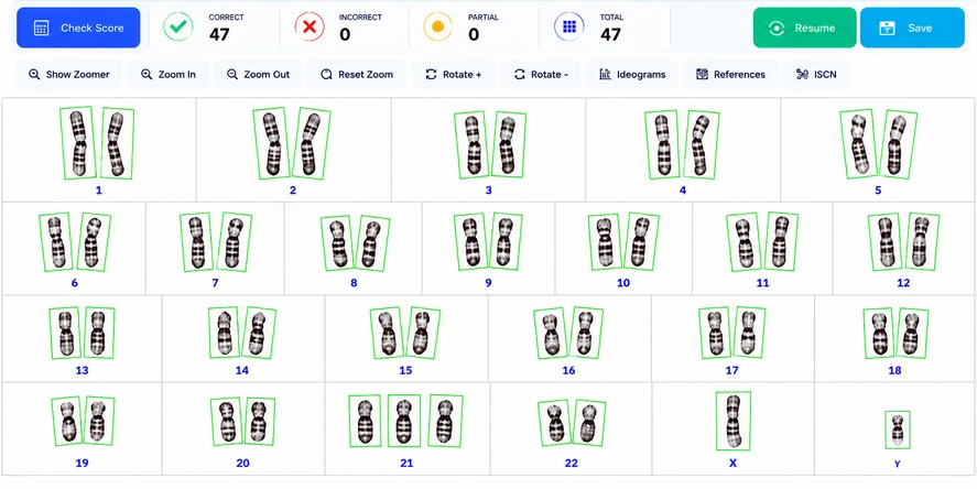
Arrange the chromosomes in the standard order, 1 to 22 followed by the sex chromosomes, with the short arm (p) at the top and the centromeres aligned. Place homologs side by side. When two homologs are at slightly different stages of condensation, match them band by band rather than by length alone.
Step 5: Analyze band by band
Compare each pair of homologs along its whole length. You are looking for:
- numerical changes: an extra chromosome (trisomy) or a missing one (monosomy);
- deletions and duplications: missing or repeated bands on one homolog;
- translocations: material exchanged between chromosomes, which changes the banding of both;
- inversions: a segment turned around within one chromosome;
- other rearrangements: isochromosomes, ring chromosomes, and marker chromosomes whose origin cannot be identified by banding.
Learn the common normal variants too. The heterochromatin blocks on 1q, 9q and 16q vary in size between people, the satellites and stalks on acrocentric chromosomes vary, and the long arm of the Y varies in length. A pericentric inversion of chromosome 9, inv(9)(p12q13), is common and generally considered a normal variant. Reporting these as abnormalities is a classic trainee error.
Step 6: Write the result in ISCN
The result is written in ISCN: the chromosome count, a comma, the sex chromosomes, then any abnormalities. A normal female is 46,XX. A male with trisomy 21 is 47,XY,+21. A female who carries a balanced reciprocal translocation between chromosomes 11 and 22 is 46,XX,t(11;22)(q23;q11.2).
See ISCN notation examples for the rules and more worked examples.
Common karyotyping mistakes
- Mixing up look-alike pairs: 4 and 5, 7 and X, the D-group chromosomes 13, 14 and 15, 19 and 20, and 21 and 22.
- Miscounting: counting a bent chromosome twice, or missing one hidden under an overlap.
- Calling a single cell a clone: random loss during slide making is common, so one abnormal cell is rarely a result.
- Reporting normal variants as abnormalities.
- Matching homologs by length only: the two homologs of a pair can differ in condensation, so compare their bands.
- Writing ISCN out of order: sex chromosome abnormalities come first, then autosomes in numerical order.
Keep learning
This page is for education and training. It is not medical advice and must not be used to diagnose or treat anyone. Laboratory results should be interpreted by qualified professionals following current standards.